|
|
|
| An Unusual Case Of Gemination, Supernumerary Tooth And Talon's Cusp. |
Ashu Gupta 1 , Anshu Minocha 2 , Vishal Sharma 3
1 MDS, Professor & Head, Department of Conservative Dentistry and Endodontics - HP Govt. Dental College and Hospital Shimla
2 MDS, Lecturer, Department of Conservative Dentistry and Endodontics - HP Govt. Dental College and Hospital Shimla
3 MDS, Asstt. Proffesor, Department of Conservative Dentistry and Endodontics - HP Govt. Dental College and Hospital Shimla
|
| Address For Correspondence |
Dr. Ashu Gupta, MDS, Professor & Head,
Department of Conservative Dentistry and
Endodontics, HP Govt. Dental College and
Hospital Shimla HP
Telephone: 9418470020
E-mail : guptaashumoni@gmail.com |
| Abstract |
| Sometimes we come across something which is unusual. The paper presents a similar unusual case of a patient with gemination, supernumerary tooth and talon's cusp in a single arch. The paper also describes the conservative way in which the treatment has been done. |
|
| Keywords |
| Gemination, Autosomal, Mesiodistal, Occlusal |
|
| Full Text |
Introduction
Gemination is a developmental anomaly of form which is recognized as an attempt by a single tooth germ to divide resulting in a large single tooth with a bifid crown and usually a common root and root canal. Primary dentition is affected more frequently than the permanent dentition, but there seems to be no gender difference. Although its prevalence is variable in different populations, it ranges from 0.1 to 1 percent. Gemination is most often seen in the maxillary incisors and canines. Both the genetic and environmental factors are believed to play a major role in the process of gemination. Spouge has suggested that the condition may result from trauma to the developing tooth bud. Case history studies have suggested that the anomaly exhibits a hereditary tendency similar to the one affecting the dental lamina and resulting in hyperdontia. The mode of inheritance appears to be either autosomal recessive or dominant with very little penetrance. Gemination or cleavage of a single tooth germ has been reported to be of two types, partial cleavage (true gemination) or complete cleavage (twinning).
The talon cusp, or dens evaginatus of anterior teeth, is a relatively rare developmental anomaly characterized by the presence of an accessory cusplike structure projecting from the cingulum area or cementoenamel junction of the maxillary or mandibular anterior teeth in both the primary and permanent dentition. The name of the anomaly is derived from the anomalous structure's resemblance to eagle's talon and it is composed of normal enamel and dentin. Most but not all talon cusps contain a pulpal extension but only a few demonstrate a visible pulpal extension on dental radiographs. Prevalence of talon cusp varies considerably in different studies in different populations and ranges from 1% to 7%. Permanent dentition and males seem to be more commonly affected. The etiology again as for gemination has been suggested to have both genetic and environmental factors.
Case report
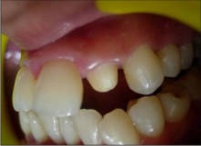
A 20 years old female patient reported in the department of operative dentistry with the chief complaint of an abnormal looking upper left lateral incisor (Fig. 1).
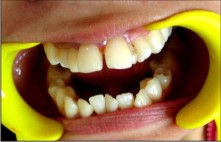 | Figure1. Preoperative photograph showing geminated maxillary left lateral incisor and supernumerary maxillary right lateral incisor.
 |
On examination, the tooth was found to be a case of gemination. The crown of the tooth had split partially into two by a clear line of demarcation in the form of a deep grove which was stained. Mesial portion of the tooth was anteriorly overlapped by the cental incisor. The width of the crown was approximately 12 mm and the mesial of the two crown structures made almost 40% of the total width of the crown of the tooth and the rest by the distal of the geminated crowns. Careful examination revealed the presence of a supernumerary right lateral incisor (Fig. 2, 3).
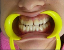
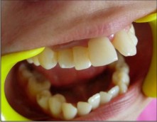 | Figure2. Preoperative photograph showing supernumerary maxillary right lateral incisor and proclined maxillary incisors.
|
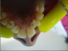
The distal right lateral incisor (there were two right lateral incisors a mesial and a distal) had a talon's cusp. The talon was conical in shape and was about 3mm in length and was interfering with the occlusion of the patient. A fissure was also noted on the lingual aspect of the mesial right lateral incisor which was in proximity to the central incisors (Fig. 3). Mild gingivitis was also noted.
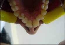 | Figure3. Occlusal photograph of the same patient taken preoperatively.
|
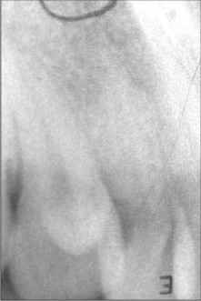
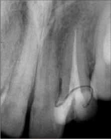
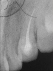
Careful history taking revealed that no one in the family of the patient had any such problem and no history of oro-facial trauma was reported by the patient. The patient was normal in all other aspects. The patient had a class I molar relation, but with increased overjet. On thermal vitality testing, all the teeth responded normally. Radiographs revealed the crowns of the left lateral incisor to have two separate pulp chambers separated by a wall of hard tooth structure and a common root and root canal (a case of true gemination), whereas on the right side both the lateral incisors had their own individual roots (a case of supernumerary tooth). The radiograph of the right lateral incisor with talon cusp gave a hint of the presence of pulp horn in the talon (Fig.4, 5).
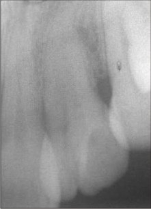 | Figure4. Preoperative radiograph of geminated maxillary left lateral incisor.
|
 | Figure5. Preoperative radiograph of supernumerary maxillary right lateral incisor with talon.
|
The situation was discussed with the patient and with an orthodontist. A treatment plan was arrived at after the discussion and described to the patient, the patient agreed to the plan.
First of all, root canal treatment was done in the left lateral incisor making two different access openings on the two geminated crowns (Fig.6, 7).
 | Figure6. Occlusal photograph of the patient showing access preparation for endodontic treatment.
|
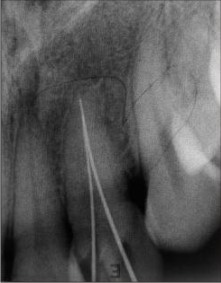 | Figure7. Working length determination of geminated maxillary left lateral incisor
|
After root canal treatment (Fig.8)
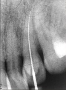 | Figure8. Working length determination of maxillary right lateral incisor with talon.
|
the pulp chambers of the tooth were cleaned and filled with light cure composite, access openings were also filled. It was decided to give a single crown of normal width on the left lateral incisor. The major problem which was anticipated was the loss of ferrule if the mesiodistal width of the crown was reduced beyond a certain point. Mesiodistally the tooth had three walls of hard structure a mesial, a central (separating the two pulp chambers), and a distal, reducing mesiodistal width involved reduction of the mesial and distal walls. Reducing any of these completely could have led to loss of ferrule. More reduction was required on the mesial wall because on mesial the tooth was overlapped by the central incisor hence, for better esthetics more reduction was needed. Tooth reduction was done very carefully to preserve the ferrule, and only to the extent the shape of the tooth permitted (Fig.11).
 | Figure11. Left lateral incisor after reduction for crown placement.
|
An impression was taken and crown was put on the tooth (Fig.12).
 | Figure12. Postoperative photograph after crown placement.
|
Root canal treatment was also done in the right lateral incisor having talon (Fig10).
 | Figure9. Radiograph showing obturation of geminated maxillary left lateral incisor.
|
 | Figure10. Radiograph showing obturation of maxillary right lateral incisor with talon.
|
After the completion of the root canal treatment, the talon was ground off and the tooth restored. Pit and fissure sealant was applied on the lingual fissure of the mesial one of the two right lateral incisors. The patient was observed for some time and then referred to an orthodontist for further treatment.
Discussion
Lot of debate has been there over terminology. Historically gemination was defined as an attempt of a single tooth bud to divide, with the resultant formation of a bifid crown and a common root and root canal but question has been raised that it may be a result of fusion of a normal tooth with the crown of a supernumerary tooth. Similarly an extra tooth if resulting from the development of two separate teeth as a result of complete separation has been referred to as twinning, it is also arguable in terms of pathogenesis. So the exact pathogenesis is questionable and may be confusing. To keep the things simple gemination is defined as a single enlarged tooth or joined i.e. double tooth crown in which the tooth count is normal when the anomalous tooth is counted as one and an extra tooth is termed as a supernumerary tooth.
As far as treatment is concerned Taylor described a case where a geminated maxillary lateral incisor which was seen as unaesthetic and treatment involved removal of the large, notched, geminated tooth and autogenous transplantation of a supenumerary lateral incisor from the opposite maxillary quadrant was done. Though our case is very similar a more conservative treatment has been done. As far as talons cusp with premature contact and occlusal interference is concerned, the talon cusp can be reduced gradually on consecutive visits at 6 to 8 weeks interval in order to allow time for deposition of reparative dentin for pulpal protection. In our case we had a different approach seeing the size of the talon and projection of pulp tissue in the talon.
Summary
Each case of dental anomaly should be seen as a different case and treatment plan should be arrived at after analyzing all the aspects related to the condition.
References
1. Mark A. Scheiner, Wayne J. Sampson Supernumerary teeth: A review of the literature andfour case reports Australian Dental Journal 1997;42:(3).
2. Neville, Damm, Allen; Oral and Maxillofacial Pathology; 2nd edition; 2004
3. Richard L. Spuller, Matthew Harrington; Gemination of a maxillary permanent central incisor treated by autogenous transplantation of a supernumerary incisor: case report, DD Pediatric Dentistry; December 1986/Vol. 8.
4. Santos, K. S. A.; Lins, C. C. S. A.; Almeida-Gomes, F.; Travassos, R. M. C. & Santos Anatomical Aspects of Permanent Geminated maxillary Central Incisors Int. J. Morphol., 27(2):515-517, 2009.
5. Shafer's Textbook of Oral Pathology 5th ed. (2006).
6. Türkaslan, Hasan Suat Gökçe, and Mehmet Dalk?z Esthetic Rehabilitation of Bilateral Geminated Teeth: A Case Report Süha European J Dent. 2007 July; 1(3): 188-191. |
|
|
|
|
|
|